Home › Without Label › Anatomy Of Body What Under Rib Age : Diaphragm Definition Function Location Britannica : Topographic anatomy and operative surgery of the abdomen.
Anatomy Of Body What Under Rib Age : Diaphragm Definition Function Location Britannica : Topographic anatomy and operative surgery of the abdomen.
Anatomy Of Body What Under Rib Age : Diaphragm Definition Function Location Britannica : Topographic anatomy and operative surgery of the abdomen.. One possible reason for pain in this area is that the colon, or large intestine, passes under the rib cage carrying faeces on its way to the. Ribs 1 2 10 11 and 12 can be described as atypic. Anatomy is the oldest scientific discipline of medicine. Related posts of rib cage organs anatomy. Skeletal systemhuman anatomy and physiologyhearthealthbronchitisdodge stealthconditions.
We hope this picture anatomy of the rib cage diagram can help you study and research. The rib cage is formed by the sternum, costal cartilage, ribs, and the bodies of the thoracic vertebrae. In most tetrapods, ribs surround the chest, enabling the lungs to expand and thus facilitate breathing by expanding the chest cavity. Learn vocabulary, terms and more with flashcards, games and other study tools. Ribs 1 2 10 11 and 12 can be described as atypic.
Diagram True Ribs Diagram Full Version Hd Quality Ribs Diagram Mapgavediagram Arebbasicilia It from www.medicalook.com We hope this picture anatomy of the rib cage diagram can help you study and research. In human anatomy, intercostal muscles (the muscles found in between your ribs) are particularly susceptible to hypoperfusion. The most superior rib is designated rib 1 and it articulates with the t1 thoracic vertebrae. The sternal angle is important in costal breathing, since it allows for greater. The body, or shaft of the rib is flat and curved. Skeletal systemhuman anatomy and physiologyhearthealthbronchitisdodge stealthconditions. The rib cage is a primarily protective structure, encircling the heart and lungs. The tenth rib attaches directly to the body of vertebra t10 instead of between vertebrae like the second through ninth ribs.
Each other and with the seventh rib.
Mainly these body planes are used in human anatomy to describe the direction and location of body structures. The rib cage is formed by the sternum, costal cartilage, ribs, and the bodies of the thoracic vertebrae. Pain rib anatomy names human ribs cages injury rib cage art rib cage and pelvis sternum anatomy diagram. Your rib bones themselves are quite fragile and are easily broken in an accident or even by a violent sneeze. True ribs (proper ribs) are directly connected to the sternum on the interior wall of the rib body is a channel, sulcus costae, with blood vessels and nerves. November 25, 2020december 3, 2019. Each other and with the seventh rib. Anatomy of a human body we study anatomy. The rib cage surrounds the lungs and the heart, serving as an important means of bony protection for. Where the manubrium articulates with the top of the body of the sternum is a sternal angle (louis' angle). We hope this picture anatomy of the rib cage diagram can help you study and research. The rib cage is a primarily protective structure, encircling the heart and lungs. Understanding the anatomy of the rib cage and its position in the body.
The internal surface of the shaft has a groove for the neurovascular supply of the the ribs are a set of twelve paired bones which form the protective 'cage' of the thorax. They articulate with the vertebral column. The rib cage surrounds the lungs and the heart, serving as an important means of bony protection for. Each pair articulates with a different thoracic vertebra on the posterior side of the body. True ribs (proper ribs) are directly connected to the sternum on the interior wall of the rib body is a channel, sulcus costae, with blood vessels and nerves.
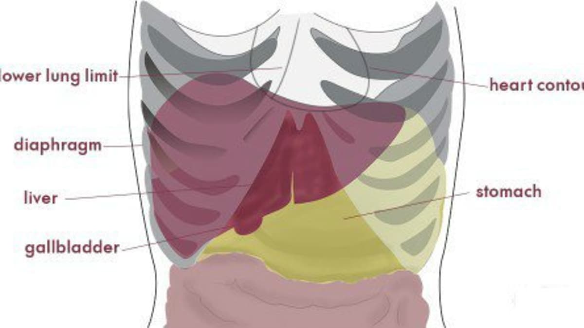
Abdominal Cavity Anatomy Britannica from cdn.britannica.com What is the term used for the articulation? Linea spinarum connects both upper anterior spines of ilium. The sternal angle is important in costal breathing, since it allows for greater. A fractured rib is very painful. In human anatomy, intercostal muscles (the muscles found in between your ribs) are particularly susceptible to hypoperfusion. True ribs (proper ribs) are directly connected to the sternum on the interior wall of the rib body is a channel, sulcus costae, with blood vessels and nerves. One possible reason for pain in this area is that the colon, or large intestine, passes under the rib cage carrying faeces on its way to the. Your ribs form a protective cage that encloses many of your delicate internal organs, such as your heart and lungs.
The muscles and the bones are under the layer of subcutaneous fat.
Pain rib anatomy names human ribs cages injury rib cage art rib cage and pelvis sternum anatomy diagram. In human anatomy, intercostal muscles (the muscles found in between your ribs) are particularly susceptible to hypoperfusion. Your rib bones themselves are quite fragile and are easily broken in an accident or even by a violent sneeze. Understanding the anatomy of the rib cage and its position in the body. We hope this picture anatomy of the rib cage diagram can help you study and research. Linea spinarum connects both upper anterior spines of ilium. The internal surface of the shaft has a groove for the neurovascular supply of the the ribs are a set of twelve paired bones which form the protective 'cage' of the thorax. Each other and with the seventh rib. Top suggestions for anatomy under rib cage. Learn vocabulary, terms and more with flashcards, games and other study tools. Important anatomical landmark located in the superior portion of the manubrium. The under surface is smooth and without a costal groove. The most superior rib is designated rib 1 and it articulates with the t1 thoracic vertebrae.
Under the left side of your rib cage are your heart your left kidney left lung and spleen. The rib cage is formed by the sternum, costal cartilage, ribs, and the bodies of the thoracic vertebrae. Understanding the anatomy of the rib cage and its position in the body. The body, or shaft of the rib is flat and curved. Linea spinarum connects both upper anterior spines of ilium.
Severe Pain On The Right Side Of The Back Abdomen And Ribs Youmemindbody from images.saymedia-content.com The thorax is anatomical structure supported by a skeletal framework (thoracic cage) and contains the the head of the rib forms the posterior end of a typical rib and articulates with the costal facet located on the body of the same numbered thoracic vertebra and. Top suggestions for anatomy under rib cage. Ribs 1 2 10 11 and 12 can be described as atypic. Mainly these body planes are used in human anatomy to describe the direction and location of body structures. Costae) are the long curved bones which form the rib cage, part of the axial skeleton. The rib cage protects the organs in the thoracic cavity, assists in respiration, and provides support for the upper extremities. In human anatomy, intercostal muscles (the muscles found in between your ribs) are particularly susceptible to hypoperfusion. The actual term derives from the greek verb anatomein, which means to cut open, to dissect.
They articulate with the vertebral column.
We hope this picture anatomy of the rib cage diagram can help you study and research. The ribs are curved, flat bones which form the majority of the thoracic cage. The rib cage is a primarily protective structure, encircling the heart and lungs. Under the left rib cage there is the left lung, scapula, ascending aorta, sternum, diaphragm, spleen its just below your rib cage on the left side of your body. The thoracic cage surrounds and protects the heart and lungs in the thoracic cavity. Where the manubrium articulates with the top of the body of the sternum is a sternal angle (louis' angle). Linea costarum connects ends of the х ribs. The first documented scientific dissections on the human body are carried out as early as the third century b.c. Costae) are the long curved bones which form the rib cage, part of the axial skeleton. November 25, 2020december 3, 2019. Under the left side of your rib cage are your heart your left kidney left lung and spleen. Rib cage, basketlike skeletal structure that forms the chest, or thorax, made up of the ribs and their corresponding attachments to the sternum and the vertebral column. Skeletal systemhuman anatomy and physiologyhearthealthbronchitisdodge stealthconditions.


Post a Comment
Post a Comment